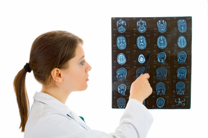
Children and young adults scanned multiple times by computed tomography (CT), a commonly used diagnostic tool, have a small increased risk of leukemia and brain tumors in the decade following their first scan, says a new study reported in the British medical journal, The Lancet.1
"This cohort study provides the first direct evidence of a link between exposure to radiation from CT and cancer risk in children," said senior investigator Amy Berrington de González, Ph.D., Division of Cancer Epidemiology and Genetics, at the National Cancer Institute. "Ours is the first population-based study to capture data on every CT scan to an individual during childhood or young adulthood and then measure the subsequent cancer risk."
The researchers emphasize that when a child suffers a major head injury or develops a life-threatening illness, the benefits of clinically appropriate CT scans should outweigh future cancer risks.
CT scan not needed for concussion
While concussions are head injuries, they are typically associated with grossly normal structural neuroimaging studies and don't show up on most magnetic resonance imaging (MRI) exams or CT scans. As a result, conventional CT or MRI scans of the brain are usually not needed where post-concussion signs or symptoms are mild and clear within a week to ten days.
A CT or MRI is, however, recommended in some circumstances, including where there is or has been:
- Loss of consciousness (LOC) for more than a minute seconds (remember: a concussions may or may not involve LOC, with 90% of all concussions occurring without LOC);
- Prolonged impairment of the conscious state, especially if there is any suggestion of a deteriorating level of consciousness;
- Dramatic worsening of a headache;
- Speech or language difficulties such as aphasia or dysarthria (impaired speech and language skills), poor enunciation, poor understanding of speech, impaired writing,impaired ability to read or to understand writing, inability to name objects (anomia)
- Vision changes such as reduced vision, decreased visual field, sudden vision loss, double vision (diplopia)
- Neglect or inattention to the surroundings on one side of the body
- Loss of coordination, or loss of fine motor controll (ability to perform complex movements)
- One-sided eyelid drooping, lack of sweating on one side of the face, and sinking of one eye into the socket
- Poor gag reflex, swallowing difficulty, and frequent choking
- Seizure activity; or
- Worsening post-concussion signs or symptoms, or persistent symptom (longer than 7 to 10 days)(e.g. post-concussion syndrome)
CT scans deliver a dose of ionizing radiation to the body part being scanned and to nearby tissues. Even at relatively low doses, ionizing radiation can break the chemical bonds in DNA, causing damage to genes that may increase a person's risk of developing cancer. Children typically face a higher risk of cancer from ionizing radiation exposure than do adults exposed to similar doses.
The investigators obtained CT examination records from radiology departments in hospitals across Britain and linked them to data on cancer diagnoses and deaths. The study included people who underwent CT scans at British National Health Service hospitals from birth to 22 years of age between 1985 and 2002. Information on cancer incidence and mortality from 1985 through 2008 was obtained from the National Health Service Central Registry, a national database of cancer registrations, deaths and emigrations.
Approximately sixty percent of the CT scans were of the head, with similar proportions in males and females. The investigators estimated cumulative doses from the CT scans received by each patient, and assessed the subsequent cancer risk for an average of 10 years after the first CT. The researchers found a clear relationship between the increase in cancer risk and increasing cumulative dose of radiation. A three-fold increase in the risk of brain tumors appeared following a cumulative absorbed dose to the head of 50 to 60 milligray (abbreviated mGy, which is a unit of estimated absorbed dose of ionizing radiation). Similarly, a three-fold increase in the risk of leukemia appeared after the same dose to bone marrow (the part of the body responsible for generating blood cells). The comparison group consisted of individuals who had cumulative doses of less than 5 mGy to the relevant regions of the body.
The absorbed dose from a CT scan depends on factors including age at exposure, sex, examination type, and year of scan. Broadly speaking, two or three CT scans of the head using current scanner settings would be required to yield a dose of 50 to 60 mGy to the brain. The same dose to bone marrow would be produced by five to 10 head CT scans, using current scanner settings for children under age 15.
In countries like the United States and Britain, the use of CT scans in children and adults has increased rapidly since their introduction 30 years ago. Due to efforts by medical societies, government regulators, and CT manufacturers, scans performed on young children in 2012 can have 50 percent lower radiation doses, compared to scans carried out in the 1980s and 1990s, say the investigators. However, the amount of radiation delivered during a single CT scan can still vary greatly and is often up to 10 times higher than that delivered in a conventional X-ray procedure.
"CT can be highly beneficial for early diagnosis, for clinical decision-making, and for saving lives. However, greater efforts should be made to ensure clinical justification and to keep doses as low as reasonably achievable," said lead author, Mark S. Pearce, Ph.D., Institute of Health and Society, Newcastle University.
Source: National Cancer Institute
1. Pearce MS, et al. Radiation exposure from CT scans in childhood and subsequent risk of leukaemia and brain tumours: a retrospective cohort study. The Lancet. June 7, 2012 (published online ahead of print).
Posted June 20, 2012


